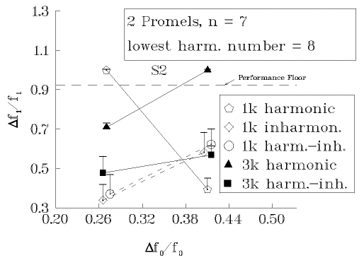
Figure 3: Results of Experiment 1 for Subject 1 (Panel a) and Subject 2 (Panel b). The component sinusoids of the two promels were harmonic in both (“harmonic”), harmonic in one and inharmonic in the other (“harm.-inh.”), or inharmonic in both promels (“inharmonic”). The lowest harmonic number in the low-fo promel was 8 only in the harmonic conditions; for the other harmonic numbers, refer to Table 1. Abscissa: normalized fundamental frequency difference of the two promels; ordinate: Weber fraction for the peak formant frequency difference.
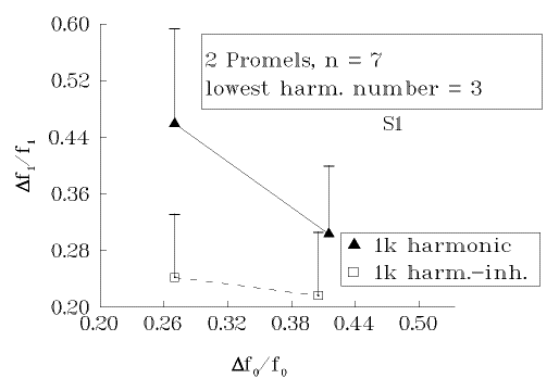
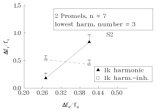
Figure 4: Results of Experiment 1 for Subject 1 (Panel a) and Subject 2 (Panel b). The component sinusoids of the two promels were harmonic in both (“harmonic”), harmonic in one and inharmonic in the other (“harm.-inh.”), or inharmonic in both promels (“inharmonic”). The lowest harmonic number in the low-fo promel was 3 only in the harmonic conditions; for the other harmonic numbers, refer to Table 1. Abscissa: normalized fundamental frequency difference of the two promels; ordinate: Weber fraction for the peak formant frequency difference.
Figure 5: Schematic diagram of the stimulus of Experiment 2 shown in the time-frequency plane. The fundamental frequency contour of the two promels were mirror images of one another, sweeping up-down-up and down-up-down, respectively, between the bottom frequency f01 and the top frequency f02 .

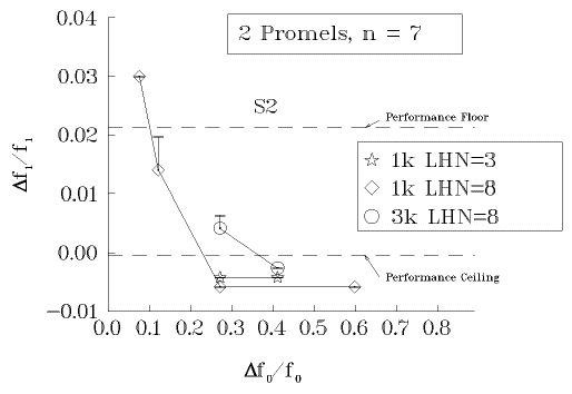
Figure 6: Results of Experiment 2 for Subject 1 (Panel a) and Subject 2 (Panel b). The component sinusoids of the two promels were always harmonic. The lowest harmonic number in the low-fo promel was 3 (“LHN=3”), or 8 (“LHN=8”); the promels' harmonic region was 1kHz or 3kHz.. Abscissa: normalized fundamental frequency difference of the two promels; ordinate: Weber fraction for the peak formant frequency difference. Data outside the lower and upper limits of the adaptive paradigm are only estimated and are indicated as being either below the “performance floor” or above the “performance ceiling”.